ABOUT MRI SCAN (MAGNETIC RESONANCE IMAGING)
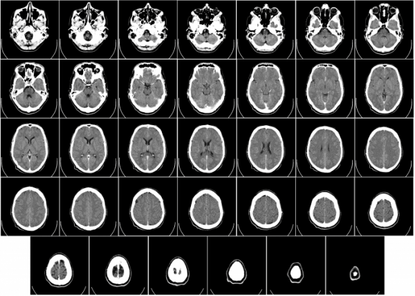
MRI (magnetic resonance imaging) scan is used to create detailed images of internal organs and tissue. The process involves the use of magnets and radio waves to create high resolution and detailed images of an organ or tissue, in order to help making a diagnosis or devising a treatment plan.
An MRI scan can be performed on the brain, chest, abdomen, heart or spine and is a painless process. Some scans may require a contrast material to be injected into the blood, in order to help create as much detail as possible.
Similar scans include CT (computerized tomography) scan and PET (positron emission tomography) scans. However, an MRI scan is most commonly performed to diagnose brain and spinal cord injuries or conditions, as it can detect a brain aneurysm, tumors and the effects of a stroke.
Recommended for
- Patients who have sustained a brain or spinal injury
- Patients with tumors
- Patients who have vascular disease, congenital heart condition, or cardiomyopathy
- Cancer patients
TIME REQUIREMENTS
- Number of days in hospital: 1 .
Patients can usually leave directly after the MRI scan is complete.
- Average length of stay abroad: 1 – 5 days.
It may take a few days for the results of the scan to be processed and patients will usually attend a follow up consultation to discuss the results.
- Number of trips abroad needed: 1.

COMPARE MRI SCAN (MAGNETIC RESONANCE IMAGING) PRICES AROUND THE WORLD
| Country | Cost |
|---|---|
| United States | 2550€ |
| Mexico | 516€ |
| Turkey | 269€ |
| Hungary | 200€ |
| Poland | 200€ |
HOW TO FIND QUALITY TREATMENT ABROAD
BEFORE MRI SCAN (MAGNETIC RESONANCE IMAGING) ABROAD
Ahead of the MRI scan, patients should advise their doctor if they are claustrophobic, as sedation can be arranged if necessary.
Patients should plan to wear comfortable clothing which do not have metal buttons or zips etc. and remove any piercings or jewelry from their body.

HOW IS IT PERFORMED
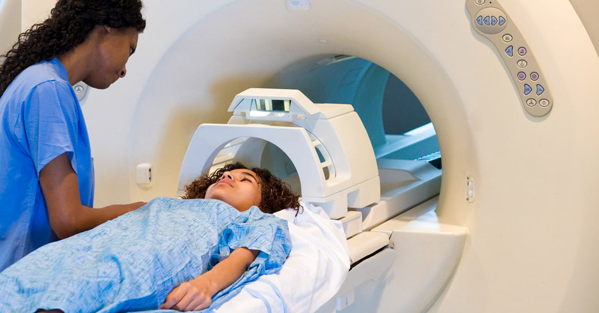
Depending on the part of the body that is being scanned, the patient may need to remove their clothing and wear a hospital gown, and contrast material may be injected into the blood to help to create detailed images. The patient will be asked to lie as still as possible on a bed which is attached to the scanning machine.
The bed will pass through the tube section of the machine which will take multiple images of the organ or tissue being scanned. Depending on the part of the body that is being scanned, the bed will pass through the tube either head or feet first.
In the case where an MRI of the brain is being taken, the patient will have a piece placed over their head to keep it in place, and it may have a mirror attached to it, so that the patient can see the radiologist in the adjoining room. There is also a microphone in the room so that the patient can communicate with the radiologist, should they experience any discomfort.
As the machine is quite loud, patients may be given headphones with music to help with the noise. Once the images are taken, they are sent to a computer to be interpreted by the radiologist. It may take a few days for the results to be processed and the patient will then need to attend a follow up consultation appointment to discuss the findings.
Materials
Contrast material may be used.
Anesthesia
Sedation may be administered to nervous or claustrophobic patients.
Procedure duration
The MRI Scan (Magnetic Resonance Imaging) takes 20 to 45 minutes.
Possible discomfort
Patients may feel uncomfortable during the scan due to the loud noises of the machine and the restriction to move.
IMPORTANT THINGS TO KNOW ABOUT MRI SCAN (MAGNETIC RESONANCE IMAGING)
Not recommended for
- Patients with pacemakers
- Pregnant patients
- Patients who have an IUD (intrauterine device)
Potential risks
- Allergic reaction to contrast material that may be used
- Exposure to small amounts of X-ray radiation
FREQUENTLY ASKED QUESTIONS
MRI stands for Magnetic Resonance Imaging. MRI scans work by using a powerful magnetic field, radio frequency pulses, and computer software to change the way atoms in your body align. As they realign, they emit energy which is different in different bodily tissues. The computer reads these energy signals and uses them to make an image for the doctor.
Patients must remain completely still during the MRI scan, which can be difficult for long periods of time. Some patients also experience claustrophobia, or find the sounds made by the machine difficult to tolerate. These patients might wear headphones and listen to music during the exam, wear earplugs, or be given a mild sedative to help them be more comfortable.
The average patient experiences almost no risk with MRI scans. If sedation is used, there is a small risk of complications involved with the sedative being used. For some scans, a contrast dye will be injected into the bloodstream, which poses a small risk of complication. Other than the injection, there is no pain associated with an MRI.
Although very low levels of radiation are used and there are no studies suggesting MRI’s may be dangerous to an unborn fetus, pregnant women are advised not to get an MRI unless it is absolutely necessary. If you are pregnant or think you might be, discuss any concerns with your doctor.
MRI scans are used to create images of soft tissues inside the body. They can be used to take images of a variety of tissues that are softer than bone, and are used in the treatment of a variety of conditions from tracking the healing of torn ligaments, to checking the progress of cancer treatment.
The accuracy of MRI results depends largely on the skill and experience of the medical professional interpreting the images.


